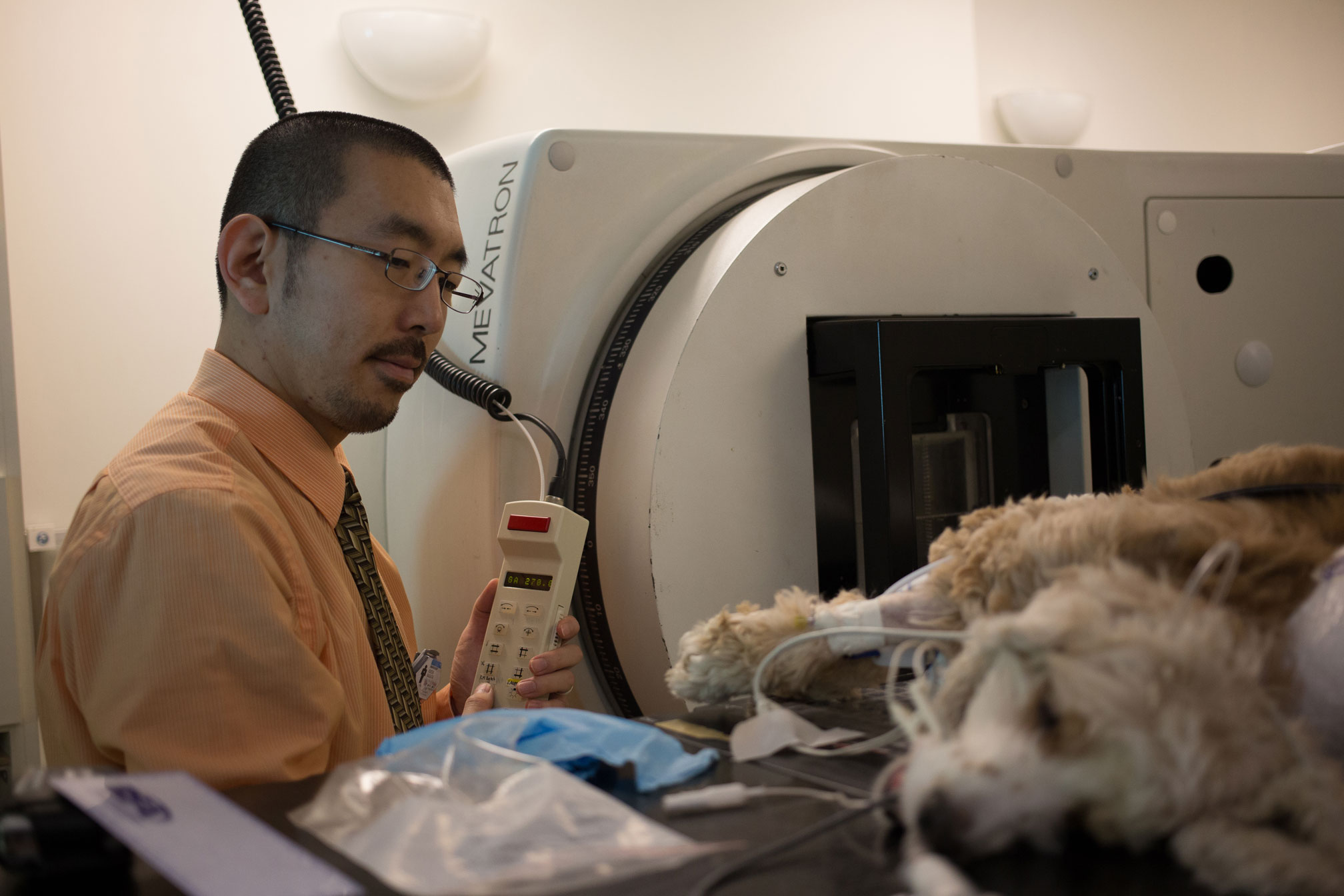
Athens, Ga. – The University of Georgia Veterinary Teaching Hospital recently added a new treatment option to its oncology service, intensity-modulated radiation therapy, and a neuronavigator to its neurology and neurosurgery service. Both the IMRT and the neuronavigator are currently available in only a handful of veterinary teaching hospitals in the U.S.
IMRT is different from traditional radiation therapy in that it allows clinicians to conform the shape of the radiation being administered to closely match the outline of the tumor being treated. This allows for a more targeted, higher dose of radiation to be delivered to the tumor, which may lead to a longer tumor-control time. More importantly, using the IMRT technique leads to a lower dose of radiation to the sensitive vital organs that surround the tumor, which results in fewer side effects.
“IMRT is especially useful when the tumor has a complicated shape and/or surrounds an animal’s sensitive organs,” said Koichi Nagata, DVM, DACVR (Radiation Oncology). “Multiple tumors that are close to each other can also be treated with a better radiation dose distribution, compared to radiation therapy without using IMRT.”
Nagata is an assistant professor of radiation oncology within the UGA College of Veterinary Medicine, and is heading up the hospital’s IMRT program.
He began administering IMRT at the hospital earlier this month, using its linear accelerator in conjunction with specialized radiation-planning computer software. This software is able to perform highly complicated calculations that are required to generate an IMRT plan.
The IMRT procedure was first used in a select few human cancer centers in the late 1990s. While it is now available in veterinary medicine as well, there are only a handful of veterinary teaching hospitals across the United States that offer it, said Nagata.
Emory donates neuronavigator to VTH
Thanks to a generous equipment donation from Emory University Hospital Midtown in Atlanta, the UGA VTH’s neurology and neurosurgery service now has a refurbished Medtronic StealthStation TREON Surgical Navigation System, or “neuronavigator.”
The system, which is about the size of a modern ultrasound unit, can be used to diagnose diseases of the brain and also to deliver therapeutic treatments to the brain.
“It maps the skull’s size and shape, then navigates within the cranial cavity much like a Global Positioning System, or GPS, would work,” explained Simon Platt, BVM&S, MRCVS, DACVIM (Neurology), DECVN, and a professor of neurology at the College.
A patient must first undergo an MRI sequence while wearing markers that collect data about that patient’s skull and brain. Later, when that patient is in front of the neuronavigator, the veterinarian points a wand toward the patient’s head, and the wand uses the information from the MRIs and the data transmitted by the markers to create a 3-D map of the patient’s skull and brain. The neuronavigator can then guide doctors to deliver therapeutics to a precise location, or to take a biopsy of an area of the brain that cannot otherwise be accessed without surgery-or, in some cases, not accessed at all due to location.
“This allows us to treat any type of brain lesion, but particularly tumors, because we can get there accurately with minimal risk to the patients,” said Platt.
Platt added that because the equipment received was designed for human patients, it will be less cumbersome to use than the veterinary neuronavigators.
“Roughly three veterinary teaching hospitals in North America currently have neuronavigators and, to the best of my knowledge, we are the only one with a model that is typically used on human patients,” said Platt.
Clinical Trials
The hospital is continuing its clinical trial to study the post-surgical delivery of Cetuximab via convection enhanced delivery for treatment of canine brain tumors, specifically gliomas. Up to 15 dogs will be accepted into the trial. All dogs will undergo surgery to remove/debulk the tumor as much as surgically possible. At the time of the surgery, a small catheter is inserted into the residual tumor, which enables the treatment to be slowly delivered to the tumor site over a 12- to 24-hour period. Follow-up MRI examinations occur at 30 days and three months after surgery. Patients must meet specific criteria, including diagnosis, for enrollment in the trial, and pet owners must consent to participation in the trial. Referral veterinarians are encouraged to refer dogs with brain disease as possible candidates for the trial.
In addition, a new clinical trial has been launched to test a hand-held device that safely stimulates the vagus nerve transcutaneously for treatment of refractory seizure activity associated with canine epilepsy. The device has been used to treat people who suffer from primary headaches and acute asthma. As part of a pilot study, the UGA VTH tested the device on eight dogs with seizure disorders; the owners of the patients did not report any side effects or safety concerns. Patients must meet specific criteria, including diagnosis, for enrollment in the trial, and pet owners must consent to participation in the trial. Once enrolled, study participation lasts 24 weeks.
For more information on these and other clinical trials, see vet.uga.edu/research/clinical/current or call the UGA VTH at 706-542-3221.
The UGA Veterinary Teaching Hospital treats more than 22,500 animals annually. 24-hour emergency services are offered at the VTH with no referral required. For more information, see http://www.vet.uga.edu/hospital. For more about the UGA College of Veterinary Medicine, founded in 1946, see www.vet.uga.edu.


